About Us

NeuroHarmoniX Labs Pvt. Ltd. builds infrastructure for quantitative neuroimaging, enabling brain MRI data to be analyzed through structured, reproducible workflows.

The platform is informed by peer-reviewed neuroimaging research published in the academic literature. The founder of NeuroHarmoniX has contributed to several of these methodologies, which form part of the scientific foundation of NeuroHL.

NeuroHL focuses on translating validated neuroimaging methods into consistent, clinically usable workflows that support reliable analysis across clinical and research environments.

About Us

Why NeuroHL exists

NeuroHarmoniX Labs was founded to address a gap in neurological care: structural MRI is routinely performed, yet quantitative analysis, such as determining whether a hippocampus falls in the 6th percentile for a 65-year-old South Asian woman, or whether temporal lobe thinning is consistent with early Alzheimer’s disease, is rarely available in clinical workflows.

The methods to perform this analysis have existed for years within academic neuroimaging labs. What has been missing is a reliable, clinical-grade framework that applies these methods to real-world data, incorporates population-relevant context, and delivers structured outputs for clinical use.

That is what NeuroHL builds.

Every output is verified through an automated quality control pipeline that evaluates label integrity, brain volume consistency, bilateral symmetry, and spatial alignment before release.

Our Vision

A future where every brain MRI is analyzed through reproducible, population-referenced pipelines enabling earlier detection, more accurate assessment, and longitudinal monitoring of neurological conditions.

Our Mission

To make quantitative brain MRI analysis accessible to neurologists and researchers across India through reproducible, clinically usable workflows.

Our Leadership

Prasanna Parvathaneni, PhD

Founder and Chief Architect

Sydulu Maheswarapu, PhD

Academic and Strategic Advisor

Mahender Bogi

Director - Operations & Delivery

Our Core Capabilities

Human brain poster illustrated silhouette of head profile with text description of glowing brain areas vector illustration

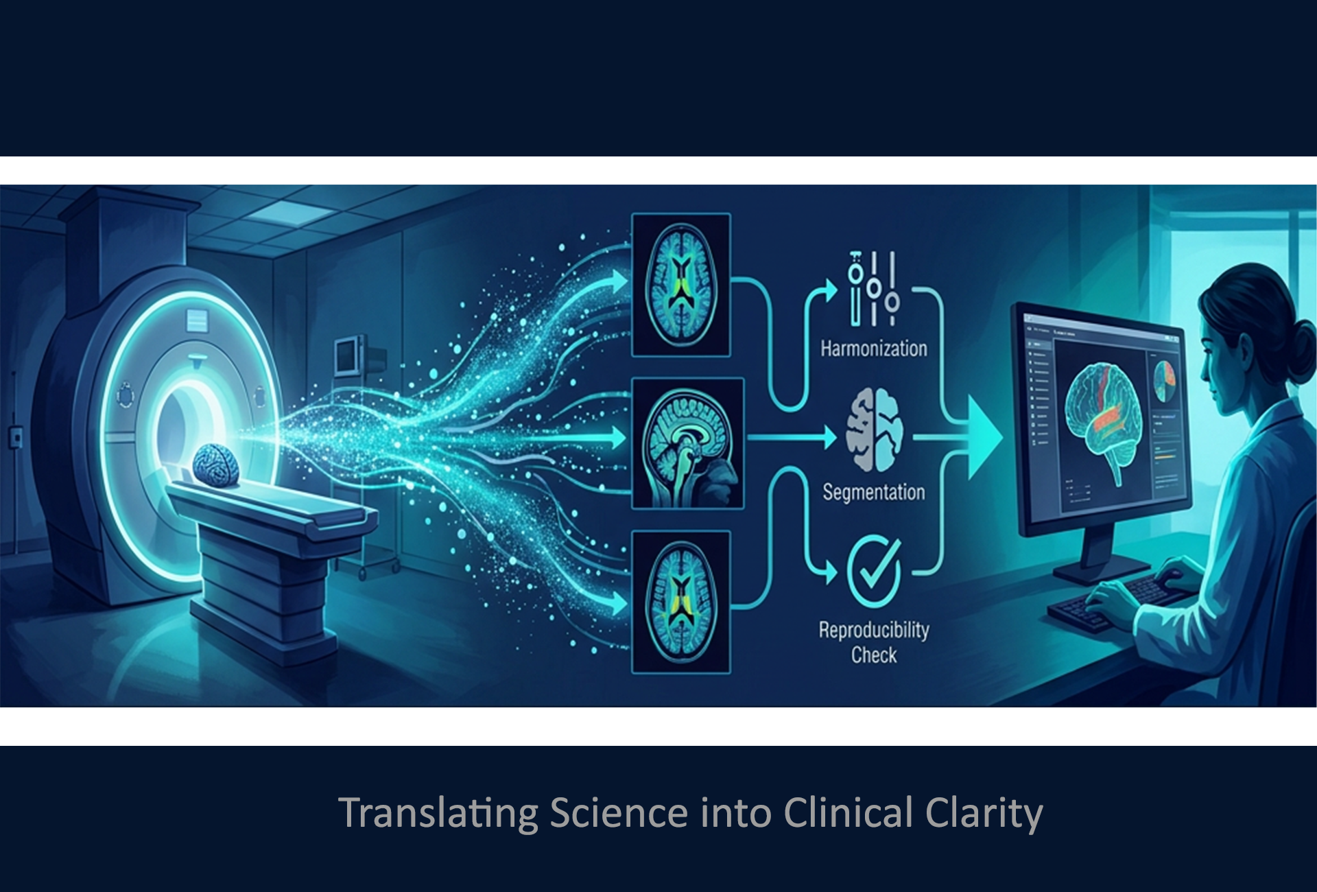
Quantitative Structural MRI

Volumetrics & Cortical Morphometry

Automated processing of 3D T1-weighted MRI scans for precise brain volumetrics, cortical thickness, and subcortical segmentation. Longitudinal tracking with normative Z-scores.
31612

Diffusion MRI Analytics

Gray & White Matter Microstructure

Advanced diffusion MRI analysis quantifying microstructural integrity across white matter tracts, cortical gray matter, and deep nuclei. FA, MD, and tract-specific metrics.
2151917428

Longitudinal Monitoring

Stable Tracking Framework

Version-controlled processing with drift detection and trajectory modeling. Reliable progression measurement distinguishing biological change from scanner variability.
5274

Cross-Scanner Harmonization

Multi-Site Standardization

Scanner-bias correction enabling multi-site dataset pooling and research consortium readiness. Harmonized metrics across vendors and field strengths..

Interested in bringing advanced MRI neuroimaging analytics to your institution?

Let’s explore a pilot or collaboration.

20+

Experience
in the Medical Image Analysis & AI Systems

We focus on:

Standardized inputs across scanners and protocols
Real-world research and clinical relevance
Automated, auditable pipelines with quality control
Interpretable quantitative outputs

Our Approach

Harmonize

Automate

Analyze

Translate

Scroll to Top